Ventrikeltachycardie: verschil tussen versies
Naar navigatie springen
Naar zoeken springen
Geen bewerkingssamenvatting |
Geen bewerkingssamenvatting |
||
| Regel 1: | Regel 1: | ||
{{Hoofdstuk|Ventriculaire ritmestoornissen}} | {{Hoofdstuk|Ventriculaire ritmestoornissen}} | ||
{{Arrhythmias| | {{Arrhythmias| | ||
| name = | | name = Ventrikeltachycardie (VT of V-tach) | ||
| locatieImage = | | locatieImage = | ||
| atrial_frequency = 60-100 | | atrial_frequency = 60-100 /min | ||
| ventricular_frequency = 110-250 | | ventricular_frequency = 110-250 /min | ||
| regularity = | | regularity = regulair | ||
| origin = | | origin = ventrikels | ||
| p_wave = AV- | | p_wave = AV-dissociatie | ||
| adenosine = | | adenosine = geen (evt. versnelling) | ||
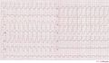
| example = | | example = Een voorbeeld van een polymorfe ventrikeltachycardie [[Image:vtach.png|250px|Ventrikeltachycardie]] | ||
| example2 = | | example2 = | ||
| animation = <flash>file=TenTusscherVT.swf|width=300|height=300|quality=best|align=right||</flash> | | animation = <flash>file=TenTusscherVT.swf|width=300|height=300|quality=best|align=right||</flash> | ||
| animationdesc = | | animationdesc = Deze animatie toont een computermodel van ventrikeltachycardie in een menselijk hart. De VT wordt geïnitialiseerd door een ventriculaire extrasystole en in stant gehouden door een re-entry rotor inferior in de linker ventrikel.<cite>tentusscher</cite> Lees [[Copyright|hier]] over het gebruik dit soort afbeeldingen in een presentatie.<br>[[Media:TenTusscherVT.swf|Link naar de file / vergroting]] | ||
}} | }} | ||
Versie van 12 aug 2007 13:25
| Dit is onderdeel van het hoofdstuk: Ventriculaire ritmestoornissen |
| {{{locatieafbeelding}}} | |
| Atriale frequentie | 60-100 /min |
| Ventriculaire frequentie | 110-250 /min |
| Regelmaat | regulair |
| Oorsprong ritme | ventrikels |
| P-top | AV-dissociatie |
| Effect van adenosine | geen (evt. versnelling) |
Voorbeeld-ecg: Een voorbeeld van een polymorfe ventrikeltachycardie 
| |
Ventricular tachycardia is defined as a sequence of three or more ventricular beats. The frequency must by higher than 100 bpm, mostly it is 110-250 bpm.
Ventricular tachycardias often origin around old scar tissue in the heart, e.g. after myocardial infarction. Also electrolyte disturbances and ischemia can cause ventricular tachycardias. The cardiac output is often strongly reduced during VT resulting in hypotension and loss of conciousness. VT is a medical emergency as it can deteriorate into Ventricular fibrillation and thus mechanical cardiac arrest.
Ventricular tachycardia can be catechorized as follows:
- Non-sustained VT: three or more ventricular beats with a maximal duration of 30 seconds.
- Sustained VT: a VT of more than 30 seconds duration (or less if treated by electrocardioversion within 30 seconds).
- Monomorphic VT: all ventricular beats have the same configuration.
- Polymorphic VT: the ventricular beats have a changing configuration. The RR interval is 180-600 ms (comparable to a heart rate of 100-333 bpm).
- Biphasic VT: a ventricular tachycardia with a QRS complex that alternates from beat to beat. Associated with digoxin intoxication.
- Voorbeeld-ECG's van ventrikeltachycardie
Ventrikeltachycardie van 140 / min met een linker bundeltakblok configuratie en linker hartas.
